|
By David E. Hansen, DVM, FAVD, DAVDC
Fellow of the academy of Veterinary Dentistry
Diplomate of the American Veterinary Dental College
There are many abnormalities that can occur in the oral cavity or with the dentition at different ages. It is important for breeders to know what normal looks like, so that at key ages they can recognize a potential problem. Many abnormalities are very easy to treat inexpensively, if they are recognized early. A mild abnormality may develop into a major problem if not promptly treated. Most can still be corrected to give the dog a comfortable and functional mouth, but it can be more difficult and expensive. This article will present the normal anatomy and terminology of the oral cavity, including the teeth.
The Normal Mouth
Dental Formulas:
It is important to know the number of teeth of both the puppy and adult dog along with the eruption times of the teeth. Puppies have a total of 28 "deciduous" (baby or immature) teeth. Looking only at the right side of the mouth, there are 3 upper and 3 lower incisors, 1 upper and 1 lower canine, and 3 upper and 3 lower premolars (pm's 2,3,4, there are no deciduous first premolars). The numbers are added together and multiplied by 2 (right and left sides) to total 28.
The formula is written as follows:
2X (3/3 i, 1/1c, 3/3pm) = 28
All of these teeth should eventually be replaced with the mature (adult, permanent) counterpart along with another upper and lower premolar and 2 upper and 3 lower molars on each side (Figure 1). This results in a total of 42 teeth as indicated by the following formula:
2X (3/3I, 1/1C, 4/4PM, 2/3M) = 42
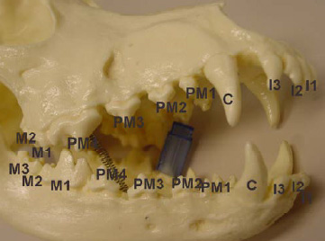
Figure 1: Adult Teeth
The times that the deciduous and mature teeth erupt are critical, as failure to erupt can cause major problems. The following chart is the normal eruption times:
Deciduous Teeth (weeks) Mature Teeth (months)
Incisors 3-4 3-5
Canines 3 4-6
Premolars 4-12 4-6
Molars 5-7
As the mature tooth bud develops, the deciduous tooth begins to have its root resorbed, resulting in exfoliation (falling off) of its crown. The exfoliation occurs as the mature tooth crown is erupting through the gingiva.
Directional terminology
Before discussion of dental anatomy, a general understanding of directional and surface terminology is required (Figure: 2). The surface of the incisor and canine teeth directed toward the lip is referred to as the "labial" surface. The surface of the premolar and molar teeth directed toward the cheek is the "buccal" surface. The surface of all of the lower (mandibular) teeth directed toward the tongue is referred to as the "lingual" surface. The surface of all of the upper (maxillary) teeth directed toward the palate is referred to as the "palatal" surface. The surface of the premolars and molars facing the teeth of the opposite jaw during closing of the mouth is called the "occlusal" surface. The term "distal" (caudal, posterior) is referring to the surface of the tooth furthest away from the middle (1st) incisor or median line. The opposite direction or surface is called "mesial" (anterior, cranial).
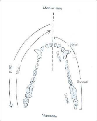
Figure 2: Directional Terminology
The Tooth
The tooth can be broken down into the crown (above the gingiva) and the root (below the gingiva)(Figure: 3). The incisor, canine, and first premolars have 1 root. The upper fourth premolars and upper first and second molars have 3 roots. The rest of the teeth have 2 roots. The "cusp" is the point or tip of the crown. The opposite end from the cusp is the "apex" or tip of the root. The term "coronal" is used to indicate the direction toward the crown tip or occlusal surface. The term "apical" is used to indicate the direction toward the root tip and away from the crown tip.
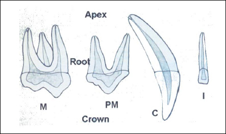
Figure 3: Parts of the Tooth
Anatomy of the Tooth
The tooth is covered by enamel on the crown and cementum on the root (Figure 4). Enamel is the hardest substance in the body. Dentin is the substance underneath the enamel and cementum. The pulp tissue is located in the pulp cavity in the central portion of the tooth. The pulp contains cells, blood vessels, lymphatics, nerves and connective tissue. The pulp functions include forming the dentin, responding to noxious stimuli, nutritional support and perception of stimuli (pain). The space where the pulp is located in the crown is called the pulp chamber and in the root is called the root canal. The apex of the tooth has multiple small openings, which allow for communication between the contents of the root canal and periapical tissues. The periodontium generally refers to the supporting apparatus of the tooth and includes the gingiva, periodontal ligament, cementum and alveolar bone. The periodontal ligament has fibers embedded in the cementum and alveolar bone, which hold the tooth in the alveolus (socket). The junction of the cementum and enamel is referred to as the cementoenamel junction (CEJ). The attachment of the gingiva to the tooth is at the level of the CEJ. In the normal healthy mouth the shallow gingival sulcus created between the tooth and the unattached gingival tissue is considered to have a normal depth of 1-3 mm in the dog.

Figure 4: Tooth Structure
Oral Cavity
The oral cavity is typically considered to be the area extending from the lips to the pharynx at the level of the tonsils. It is bound by the lips anteriorly, the cheeks laterally, by the hard and soft palate above, and by the floor of the mouth below. The pharynx is the location were the digestive and respiratory system share a common pathway. The structures, besides the teeth, located in the oral cavity are the hard and soft palate, tongue and floor of the mouth. The hard palate is the soft tissue covered, boney vault of the oral cavity. The soft tissue has a median ridge dividing the right and left side. There are transverse ridges called rugae that radiate out from the median ridge. The rugae should meet symmetrically at the median ridge. The soft palate is the unsupported soft tissue that extends back from the hard palate. The hard and soft palates serve to separate the oral cavity from the nasal cavity.
Osseous (bone) Structures
The skull and mandible support all of the teeth. The incisive bone (formerly called the premaxilla), maxilla, and mandibular bones have sockets in which the teeth are seated. The incisive bone accommodates the six upper incisor teeth. The maxilla supports the remaining of the upper teeth including the canine, four premolars and two molars in each maxilla. The mandible consists of two bilateral bones that are attached anteriorly at the midline by a strong fibrous joint called the mandibular symphysis. The mandibular bone supports the lower teeth.
Normal Occlusion
To evaluate occlusions, a basic understanding of the establishment of norms for breeds is necessary. There are three basic skull and jaw types that may be either breed or individual specific. "Dolichocephalics" are individuals with long narrow facial profiles, such as collies and greyhounds. "Brachycephalics" have a short broad facial profile, such as boxers and bulldogs. "Mesocephalics" have a more balanced facial profile that is between the other two types, such as the beagle and German shepherd.
We will concern ourselves with primarily the collie in this discussion. A dog's normal occlusion is identified by several factors. The upper incisors should overlap the lower incisors. The lower incisal edges rest on or near the lingual surface of the upper incisors in a scissors bite. The lower canine teeth are positioned midway between the upper third incisors and upper canine teeth when the mouth is shut. The lower premolar cusps should point to the inter-proximal spaces of the upper premolars in a pinking shear fashion, with the cusps of the upper fourth premolars buccal to the lower first molars. (Figure: 5)
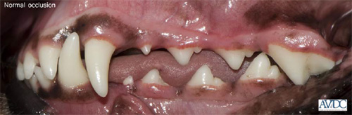
Figure 5: Normal Occlusion
|